

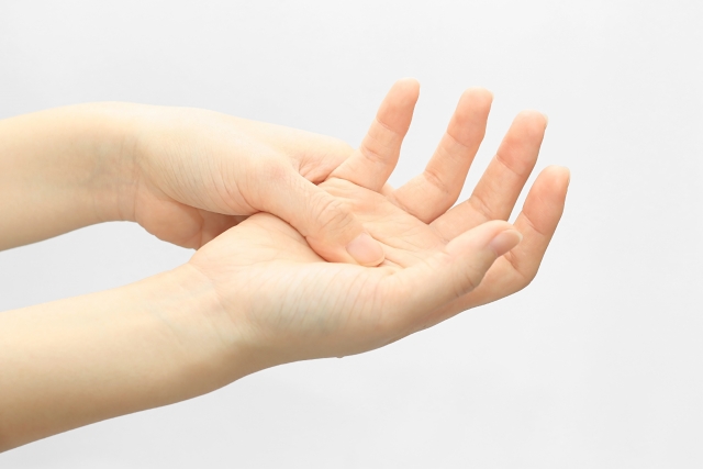
Trigger finger is a common hand problem. Pulley system is a tubular soft tissue complex over hand digits. It wraps around the flexor tendons and provides stability and lubrication upon grinding of the tendons. Overuse of the tendons or chronic repetitive action of hand digits could lead to thickening of the pulley. In severe case, this could cause compression on the flexor tendons, resulting in edema, thickening and inflammation at the tendons. Subsequently, this could limit the movement of hand digits.
The most common cause of trigger finger is overuse. Risk factors include long term carrying of heavy objects, doing housework, frequent use of computer keyboard or mobile phones. In some rare cases, trigger finger can be an initial sign of rheumatoid arthritis.
In early stage of trigger finger, there may be slow finger movement, pain and triggering during finger flexion and extension, finger joints pain. If left untreated, the condition may get worse. Patients may only be able to move their fingers by manual passively. There may also be flexion contracture of hand digits.
Oral anti-inflammatory medication or local injection of steroid can reduce the pain and relieve the triggering. Physiotherapy can be done for mobilization training. If there is no improvement by conservative treatment, operation for trigger finger release should be considered.
* Please consult doctors for further details.
Rheumatoid arthritis is an autoimmune disorder. It is caused by the dysfunction of the immune system, which wrongly attack the normal joints. This would cause inflammation of joints, resulting in redness, swelling, pain, warmth and stiffness. In severe case, there may be joint deformity. Besides joints, the disease may affect other body parts, e.g. eyes, skin, heart, cerebral vessels etc. Development of rheumatoid arthritis is related to genetic, hormonal and environmental factors.
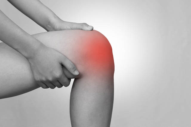
To distinguish rheumatoid arthritis from other kinds of arthritis, patients with rheumatoid arthritis may have ‘early morning stiffness’, usually more than 30 minutes. In early stage, small joints like finger, wrist or toe joints are mostly affected. In later stage, elbow, shoulder, hip, knee and ankle joints may be involved. The joints involvement is usually symmetrical, which occurs on both left and right side.
Combination of clinical condition, blood test and imaging result is required for diagnosis of rheumatoid arthritis. Clinically, position and numbers of joints with symptoms more than 6 weeks should be assessed. For blood test, rheumatoid factor (RF), Anti-cyclic citrullinated peptide (anti-CCP) antibody, C reactive protein (CRP) and erythrocyte sedimentation rate (ESR) are usually checked.
Physiotherapy may be useful to relief symptoms. Anti-inflammatory medication, disease-modifying antirheumatic drugs (DMARDS), steroid, target therapy could be used to control the disease.
* Please consult doctors for further details.
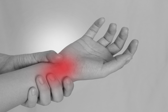
There is a tunnel bounded by carpal bones and the transverse carpal ligament at our wrist. It is called ‘carpal tunnel’. Inside the tunnel, there are median nerve and 9 flexor tendons. Carpal tunnel syndrome is a common condition usually seen in patients with chronic computer/ mouse use.
Other causes include old wrist fracture, repetitive usage of wrist, wrist deformity, diabetes mellitus, rheumatoid arthritis, renal diseases, thyroid dysfunction etc. Anything causing decrease in space of the carpal tunnel could lead to compression on the median nerve. Early symptoms include finger numbness or pain (thumb, index, middle and half of the ring finger). Muscle wasting could be seen in severe case. This could cause impairment of hand function.
With mild symptoms, patients could try resting or stretching exercise. With moderate symptoms, oral anti-inflammatory medication, drugs for neuropathic pain, local steroid injection or wrist splint could be used. If there is no improvement after conservative treatment, operation for carpal tunnel release should be considered. Nerve conduction study could be done pre-operatively for assessment to stage the disease objectively.
* Please consult doctors for further details.

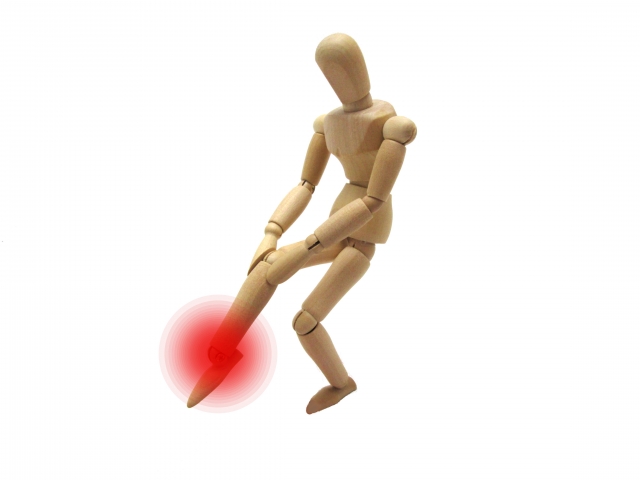
Tennis elbow is also called lateral epicondylitis. It refers to inflammation of the extensor origin of forearm muscles. It is caused by chronic overuse of forearm muscles.
Since the condition was initially discovered on tennis player, it was named as tennis elbow. However, in fact, majority of cases were not found on tennis player. Tennis elbow is commonly seen on housewife, because domestic work always involves wrist extension and contraction of forearm muscles. Long term use of computer or mobile phone increases the risk of tennis elbow.
Gout may occur when there is excessive uric acid in our body. The excessive urate crystal can stay in our joints causing inflammation. Males more than 40 years old, positive family history, obesity and menopause are the risk factors.
Purine taken in our daily diet could turns to uric acid after metabolism. The metabolism can be affected by genetic factor. Patients with high purine intake and excessive production of uric acid could aggravate gouty attack.
During acute attack, there is usually involvement of single joint (commonly the 1st metatarsophalangeal joint). There would be redness, swelling, warmth and pain. If the disease is not controlled well, there may be multiple joints involvement with frequent attack. In severe case, there may be joint deformity. In chronic gout, there may be renal function impairment or renal stone.
To prevent recurrence of gouty attack, patient should
(1) drink enough water to ease excretion of excessive uric acid
(2) avoid obesity
(3) comply with low purine diet, avoid following food
-wine
-animal internal organ
-seafood e.g. sardine fish, fish eggs, shellfish
-excessive meat
-excessive beans, mushroom, cauliflower, spinach, asparagus etc
* Please consult doctors for further details.
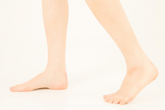
The concave surface over our medial foot is called foot arch. It composes of muscle, ligaments and bones. It provides support to our body weight, maintains balance and offers shock absorption during walking.
Flat foot refers to lowering or flattening of foot arch. More severe wear and tear could be noticed over the medial aspect at the bottom of shoes in patients with flat foot. Patient would experience fatigue over medial foot after exercise. There may be associated pain over calves, knee, hip or lumbar spine. Flat foot may worsen plantar fasciitis.
Regular exercise to strengthen foot muscle could prevent development of flat foot, e.g. tip toe stand, grasping towel with toes. Appropriate insole with arch support is also helpful.
* Please consult doctor for further details.
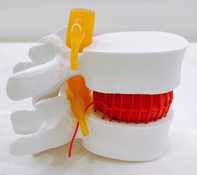
The major functions of spine include supporting body, protecting nervous system and controlling activities. There are totally 33 vertebrae in human spine. In between the vertebra, there is a soft tissue structure called intervertebral disc, which involves in shock absorption.
The central nervous system may be compressed when there is prolapsed intervertebral disc. Motor or sensory deficit of different area may be present according to the level of compression. For example, compression at cervical level could affect head, neck and upper limb; compression at lumbar level could affect buttock and lower limbs. Patients may experience weakness, numbness or pain. In severe case, there may be urinary or fecal incontinence.
Magnetic Resonance Imaging (MRI) could help to visualise the condition of prolapsed intervertebral disc and formulate treatment plan.
Physiotherapy may be able to relief the symptoms. Surgery could be considered if there is no improvement after conservative treatment.
* Please consult doctors for more details.